
Coccyx
The coccyx (pl.: coccyges or coccyxes), commonly referred to as the tailbone, is the final segment of the vertebral column in all apes,[1] and analogous structures in certain other mammals such as horses. In tailless primates (e.g. humans and other great apes) since Nacholapithecus (a Miocenehominoid),[2][3] the coccyx is the remnant of a vestigial tail. In animals with bony tails, it is known as tailhead or dock, in bird anatomy as tailfan. It comprises three to five separate or fused coccygeal vertebrae below the sacrum, attached to the sacrum by a fibrocartilaginousjoint, the sacrococcygeal symphysis, which permits limited movement between the sacrum and the coccyx.
Structure

The coccyx is formed of three, four or five rudimentary vertebrae. It articulates superiorly with the sacrum. In each of the first three segments may be traced a rudimentary body and articular and transverse processes; the last piece (sometimes the third) is a mere nodule of bone. The transverse processes are most prominent and noticeable on the first coccygeal segment. All the segments lack pedicles, laminae and spinous processes. The first segment is the largest; it resembles the lowest sacral vertebra, and often exists as a separate piece; the remaining ones diminish in size caudally.
Most anatomy books incorrectly state that the coccyx is normally fused in adults. It has been shown that the coccyx may, in some people, consist of up to five separate bony segments, the most common configuration being two or three segments.[4]
Surfaces
السطح الأمامي مقعر قليلاً، ويتميز بثلاثة أخاديد عرضية تُشير إلى نقاط التقاء القطع المختلفة. وهو يُوفر نقطة ارتكاز للرباط العجزي العصعصي الأمامي وعضلات رافعة الشرج، كما يدعم جزءًا من المستقيم . أما السطح الخلفي فهو محدب، ويتميز بأخاديد عرضية مماثلة لتلك الموجودة على السطح الأمامي، ويحتوي على صف خطي من الحديبات على كلا الجانبين - وهي النتوءات المفصلية غير المتطورة للفقرات العصعصية. من بين هذه الحديبات، يُعد الزوج العلوي هو الأكبر، ويُطلق عليه قرون العصعص، حيث يمتد باتجاه الذيل ، ويتمفصل مع قرون العجز، ويُكمل على كلا الجانبين الثقبة اللازمة لانتقال الفرع الخلفي للعصب العجزي الخامس .
الحدود
الحواف الجانبية رقيقة وتظهر عليها سلسلة من النتوءات العظمية الصغيرة، والتي تمثل النتوءات المستعرضة للفقرات العصعصية. أول هذه النتوءات هو الأكبر؛ وهو مسطح من الأمام، وغالبًا ما يمتد ليلتقي بالجزء السفلي من الحافة الجانبية الرقيقة للعجز، مُكملاً بذلك الثقبة اللازمة لانتقال الفرع الأمامي للعصب العجزي الخامس؛ أما النتوءات الأخرى فتتناقص في الحجم باتجاه الذيل، وغالبًا ما تكون غائبة. حواف العصعص ضيقة، وتُتيح اتصالًا من كلا الجانبين بالرباط العجزي الحدبي والرباط العجزي الشوكي ، وبالعضلة العصعصية والعضلة الرافعة للشرج أمام الأربطة، وبالعضلة الألوية الكبرى خلفها.
قمة
القمة مستديرة، ويرتبط بها وتر العضلة العاصرة الشرجية الخارجية ؛ ويمكن تقسيمها إلى قسمين.
الحفرة العصعصية
الحفرة العصعصية هي انخفاض سطحي على السطح الخلفي بين العجز والعجان ، وتقع في الشق بين الأليتين الذي يمتد من أسفل العجز مباشرةً إلى العجان . [ 5 ] وهي ليست موجودة بشكل ثابت لدى جميع البشر. تُشير الحفرة العصعصية إلى أعمق جزء من قاع الحوض ، بجوار العصعص. وينشأ من هذه الحفرة العضلة الرافعة للشرج . [ 6 ]
العضلة الباسطة للعصعص
The extensor coccygis is a slender muscle fascicle, which is not always present. It extends over the caudal portion of the posterior surface of the sacrum and coccyx. It arises by tendinous fibers from the last segment of the sacrum, or first piece of the coccyx, and passes downward to be inserted into the lower part of the coccyx. It is an evolutionary relic of the extensor muscle of the caudal vertebrae of other animals, enabling limited coccygeal motion.
Sacrococcygeal and intercoccygeal joints
The joints are variable and may be: (1) synovial joints; (2) thin discs of fibrocartilage; (3) intermediate between these two; (4) ossified.[7][8]
Attachments
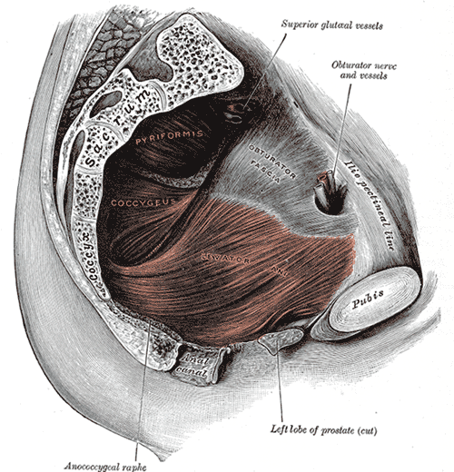
The anterior side of the coccyx has attachments to the levator ani muscle, coccygeus, iliococcygeus, and pubococcygeus, anococcygeal raphe. Attached to the posterior side is the gluteus maximus, which extends the thigh at the hip joint.[9] The ligaments attached to the coccyx include the anterior and posterior sacrococcygeal ligaments which are the continuations of the anterior and posterior longitudinal ligaments that extend along the entire spine.[9] The lateral sacrococcygeal ligaments complete the foramina for the last sacral nerve.[10] Some fibers of the sacrospinous and sacrotuberous ligaments (arising from the spine of the ischium and the ischial tuberosity respectively) also attach to the coccyx.[9] An extension of the pia mater, the filum terminale, extends from the apex of the conus, and inserts on the coccyx.
Function
The coccyx is not entirely useless in humans,[11] because it has attachments to various muscles, tendons and ligaments. However, these muscles, tendons and ligaments are also attached at many other points, to stronger structures than the coccyx. It is doubtful that the coccyx attachments are important to the well-being of humans, given the large number of coccygectomy procedures performed annually to treat coccydynia. Reviews of studies covering more than 700 coccygectomies found the operation was successful in relieving pain in 84% of cases.[12][13] 12% of the time, the only major complication faced was infection due to the proximity to the anus. One notable complication of coccygectomy is an increased risk of perineal hernia.
Clinical significance
Injuring the coccyx can give rise to a painful condition called coccydynia and one or more of the bones or the connections thereof may be broken, fractured tailbone.[14][15] A number of tumors are known to involve the coccyx; of these, the most common is sacrococcygeal teratoma. Both coccydynia and coccygeal tumors may require surgical removal of the coccyx (coccygectomy). One very rare complication of coccygectomy is a type of perineal hernia known as a coccygeal hernia.[16]
Etymology
The term coccyx is derived from the ancient Greek word κόκκυξ[17][18]kokkyx "cuckoo";[19] the latter is attested in the writings of the Greek physicianHerophilus to denote the end of the vertebral column.[20] This Greek name for the cuckoo was applied as the last three or four bones of the coccyx resemble the beak of this bird,[17][20][21][22] when viewed from the side.[9][23]
يمكن إيجاد هذا التفسير الاشتقاقي الراسخ أيضًا في كتابات عالم التشريح أندرياس فيزاليوس، الذي عاش في القرن السادس عشر، حيث كتب: os cuculi, a similitudine rostri cuculi avis [ 20 ] (يُظهر عظم الوقواق تشابهًا مع منقار طائر الوقواق). استخدم فيزاليوس التعبير اللاتيني os cuculi ، حيث os تعني عظم [ 24 ] و cuculus هو الاسم اللاتيني للوقواق. [ 24 ] يقدم عالم التشريح الفرنسي جان ريولان الأصغر، الذي عاش في القرنين السادس عشر والسابع عشر، تفسيرًا لغويًا طريفًا، إذ كتب: quia crepitus, qui per sedimentum exeunt, ad is os allisi, cuculi vocis similitudinem effingunt [ 20 ] (لأن صوت الريح الخارجة من الشرج والتي تصطدم بهذا العظم، يشبه صوت طائر الوقواق). ولا يُعتبر تفسير ريولان مقنعًا. [ 20 ] [ 21 ]
إلى جانب عظم العصعص (os cuculi) ، وعظم الذيل (os caudae) ، [ 20 ] [ 25 ] ، وُثِّق استخدام كلمة caudae للإشارة إلى الذيل [ 24 ] . قد يكون هذا التعبير اللاتيني مصدرًا للمصطلحات الإنجليزية والفرنسية والألمانية والهولندية : عظم الذيل (tailbone ) ، وعظم الذيل ( l'os de la queue) ، [ 25 ] وعظم العصعص (Schwanzbein) ، [ 21 ] [ 25 ] وعظم العصعص (staartbeen) . [ 26 ] في التسمية التشريحية اللاتينية الرسمية الحالية، Terminologia Anatomica ، [ 27 ] يُستخدم مصطلحا العصعص (coccyx) وعظم العصعص (os coccygis) .
صور إضافية
 يقع العصعص أسفل العجز وخلف تجويف الحوض .
انظر أيضاً
مراجع
- ↑ وايزبرغر، ميندي (23 مارس 2024). "لماذا لا يمتلك البشر ذيولًا؟ العلماء يجدون الإجابات في مكان غير متوقع" . سي إن إن . تم الاطلاع عليه في 24 مارس 2024 .
{{cite news}}: CS1 maint: deprecated archiveal service ( link ) - ↑ ناكاتسوكاسا 2004، اكتساب المشي على قدمين (انظر الشكل 5 بعنوان الفقرة العصعصية/الذيلية الأولى في الرئيسيات قصيرة الذيل أو عديمة الذيل. .)
- ملاحظة : يُعدّ كلٌّ من Nacholapithecus وNakaliphitecus nakayamai نوعين مختلفين من أشباه البشر في العصر الميوسيني (عينات من ناكالي وناتشولا على التوالي). انظر على سبيل المثال "مقارنات مع أشباه البشر الأخرى" في (كونيماتسو، ناكاتسوكاسا وآخرون، ديسمبر 2007).
- ↑ تيتيكر هـ، كوشار م.إ، تشولو ن، كانبيك يو، أوتاغ إ، تاشتيمور ي (2017). "تقييم تفصيلي قائم على التصوير بالرنين المغناطيسي لتشريح عظم العصعص لدى البالغين الأتراك" . المجلة النيجيرية للممارسة السريرية . 20 (2): 136-142 . doi : 10.4103/1119-3077.198313 . PMID 28091426 .
- ↑ كوزمو، ل. (2017). "سد الفجوة: حول الظواهر الطوبولوجية الجزئية الجديدة والطوبولوجيات الجزئية" . بحوث الصحة . 1 (1): 39-49 . doi : 10.31058/j.hr.2017.11004 .
- ↑ ليرس، فيرنر (2012-12-06). التشريح التطبيقي للحوض . سبرينغر. ص 40. ISBN 978-3-642-71368-2.
- ^ مين جي واي. موليني الخامس . فوترل ب (1992). "تشريح الأقراص العصعصية". مجلة طب العظام . 28 : 34 – 35.
- ↑ سالوجا ، بي جي (1988). "نسبة حدوث تكلس المفصل العجزي العصعصي" . مجلة التشريح . 156 : 11-15 . PMC 1261909. PMID 3138225 .
- 1 2 3 4 فوي، باتريك م . بوتاسي ، تشارلز جيه (3 يونيو 2008). "ألم العصعص" . الطب الإلكتروني .
- ↑ موريس، كريج إي. (2005). متلازمات أسفل الظهر: الإدارة السريرية المتكاملة . ماكجرو هيل. ص 59. ISBN 978-0-07-137472-9.
- ↑ سالادين، كينيث س. (2003). علم التشريح وعلم وظائف الأعضاء: وحدة الشكل والوظيفة ( الطبعة الثالثة). ماكجرو هيل. ص 268.
- ↑ يمكن أن يكون استئصال العصعص خيارًا علاجيًا لألم العصعص المزمن (Ugeskr Læger 2011 14 فبراير؛ 173(7): 495-500. باللغة الدنماركية. آربي، نانيت سكيليروب (1)، تروليجارد، أنطون ميتشل (2) وهيلبرج، ستين (2) https://www.coccyx.org/medabs/aarby.htm
- ↑ هيوم داي كوون وآخرون، ألم العصعص واستئصال العصعص. المجلة الكورية للعمود الفقري , 9, 4 (2012), 326-333.
- ↑ ماين، جيه واي؛ دورسونيان، إل؛ شاتيليه، جي (2000). " أسباب وآليات ألم العصعص الشائع. العمود الفقري" . العمود الفقري . 25 (23). coccyx.org: 3072–3079 . doi : 10.1097/00007632-200012010-00015 . PMID 11145819. S2CID 25790826 .
- ↑ فوي، ب.، بوتاتشي، ج.، ستيتيك، ت.، يونكلاس، ب. (2006). "حقنة ناجحة لألم العصعص" . المجلة الأمريكية للطب الفيزيائي وإعادة التأهيل . 85 (9): 783-784 . doi : 10.1097/01.phm.0000233174.86070.63 . PMID 16924191 .
- ↑ ميراندا إي بي، أندرسون إيه إل، دوسانج إيه إس، لي سي كيه (سبتمبر 2007). "الإدارة الناجحة للفتق العصعصي المتكرر باستخدام السديلة العضلية الجلدية للبطن المستقيمة منزوعة الظهارة". مجلة الجراحة التجميلية والترميمية . 62 (1): 98-101 . doi : 10.1016/j.bjps.2007.08.002 . PMID 17889632 .
- 1 2 كلاين، إي. (1971). قاموس شامل لأصول الكلمات في اللغة الإنجليزية. يتناول أصل الكلمات وتطور معانيها، موضحًا بذلك تاريخ الحضارة والثقافة. أمستردام: إلسيفير ساينس بي في
- ↑ هاربر، دوغلاس. "العصعص" . قاموس أصل الكلمات على الإنترنت .
- ↑ κόκκυξ . ليدل، هنري جورج ؛ سكوت، روبرت ؛ معجم يوناني-إنجليزي في مشروع بيرسيوس .
- 1 2 3 4 5 6 هيرتل، ج. (1880). علم التشريح التشريحي. Geschichte und Kritik der Anatomischen Sprache der Gegenwart. فيينا: فيلهلم براومولر. KK Hof- وUniversitätsbuchhändler.
- 1 2 3 كراوس، لوس أنجلوس (1844). Kritisch-etymologisches medicinisches Lexikon (Dritte Auflage). غوتنغن: Verlag der Deuerlich- und Dieterichschen Buchhandlung.
- ↑ بانورياس، آي جي؛ سترانجاليس، جي؛ ستافرينو، إل سي؛ ساكاس، دي إي (2011). "الأصول اليونانية والأبقراطية لمصطلحات العمود الفقري". مجلة تاريخ علم الأعصاب . 20 (3): 177-187 . doi : 10.1080/0964704X.2010.510180 . PMID 21736439. S2CID 22256856 .
- ↑ شوغر، أوسكار (فبراير 1995). "منظور تاريخي: عظم العصعص: العظم الذي سُمّي على اسم طائر". مجلة العمود الفقري . 20 (3): 379-383 . doi : 10.1097/00007632-199502000-00024 . ISSN 0362-2436 . PMID 7732478 .
- 1 2 3 os , cuculus , cauda . تشارلتون تي. لويس وتشارلز شورت. قاموس لاتيني ضمن مشروع برسيوس .
- 1 2 3 شريجر، CHTh.(1805). مرادف تشريحي. Synonymik der anatomischen Nomenclatur. فورث: im Bureau für Literatur.
- ^ ايفردينجن، جي جي فان، إرينبيمت، AMM فان دن (2012). Pinkhof Geneeskundig woordenboek (12de druk). هوتن: بون ستافلو فان لوغوم.
- ↑ اللجنة الفيدرالية للمصطلحات التشريحية (FCAT) (1998). المصطلحات التشريحية . شتوتغارت: ثيمي
للمزيد من القراءة
- كونيماتسو، يوتاكا؛ ناكاتسوكاسا، ماساتو؛ وآخرون . (ديسمبر 2007). "قرد عظيم جديد من العصر الميوسيني المتأخر من كينيا وآثاره على أصول القردة العليا الأفريقية والبشر" . وقائع الأكاديمية الوطنية للعلوم . 104 ( 49): 19220-19225 . رمز Bibcode : 2007PNAS..10419220K . doi : 10.1073/pnas.0706190104 . PMC 2148271. PMID 18024593 .
- ناكاتسوكاسا، ماساتو (مايو 2004). "اكتساب المشي على قدمين: سجل أشباه البشر في العصر الميوسيني ونظائره الحديثة لأشباه البشر الأوائل ثنائية القدم" . مجلة التشريح . 204 (5): 385-402 . doi : 10.1111/j.0021-8782.2004.00290.x . PMC 1571308. PMID 15198702 .
روابط خارجية
- ألم العصعص (ألم العصعص، ألم عظم الذنب) في موقع eMedicine (فصل طبي خاضع لمراجعة الأقران، متاح مجاناً عبر الإنترنت)
- عظام الصدر
- عظام العمود الفقري
- الأعضاء الأثرية