
تاج (سن)

في طب الأسنان، يُعدّ تاج السن الجزء المرئي منه فوق اللثة ، وهو عنصر أساسي في بنية السن. يُغطّى التاج بالمينا ، ويلعب دورًا حيويًا في تقطيع الطعام وطحنه. يختلف شكله وبنيته تبعًا لنوع السن ووظيفته ( القواطع ، الأنياب ، الضواحك ، أو الأضراس )، كما يختلف بين الأسنان اللبنية والأسنان الدائمة . يُساهم التاج أيضًا في جمال الوجه، والنطق، وصحة الفم.
التاج التشريحي مقابل التاج السريري
يشير التاج التشريحي إلى الجزء من السن المغطى بالمينا، سواءً كان مرئيًا أم لا. أما التاج السريري فهو الجزء الظاهر من السن داخل الفم. في البالغين الأصحاء، تتبع اللثة عادةً محيط التقاء المينا بالجذر، لذا يكون حجم التيجان السريرية والتشريحية متقاربًا. مع ذلك، قد يتغير هذا مع التقدم في السن أو الإصابة بأمراض اللثة . [ 1 ]
مصطلحات أسطح الأسنان
يستخدم أخصائيو طب الأسنان عدة مصطلحات قياسية لوصف موقع واتجاه أسطح التاج.
يُطلق على سطح السن المواجه للشفاه أو الخدين اسم السطح الوجهي. في الأسنان الأمامية، مثل القواطع والأنياب، يُعرف هذا السطح تحديدًا بالسطح الشفوي، بينما في الأسنان الخلفية، مثل الضواحك والأضراس، يُطلق عليه اسم السطح الخدي. [ 1 ]
السطح اللساني هو جانب السن المواجه للسان. في الفك العلوي أو القوس الفكي العلوي، قد يُشار إلى هذا السطح أيضًا بالسطح الحنكي نظرًا لقربه من الحنك. [ 1 ]
السطح الإطباقي هو سطح المضغ الموجود على الأسنان الخلفية (الضواحك والأضراس)، بينما تحتوي الأسنان الأمامية على حافة قاطعة، وهي حافة حادة تُستخدم للعض. [ 1 ]
The sides of a tooth that make contact with neighbouring teeth are called proximal surfaces. If the surface faces toward the midline of the dental arch, it is known as the mesial surface. Conversely, if it faces away from the midline, it is termed the distal surface.[1]
Function of the tooth crown
The crown contributes to multiple functions, including mastication, speech, aesthetics, and protection of supporting oral structures. Incisors, positioned at the front of the mouth, have sharp edges for cutting food and aiding in speech. Canines have pointed cusps to tear food and support the bite. Premolars combine tearing and grinding functions, while molars, with their broad surfaces, are specialised for crushing and grinding food.[2]
Structure of the anatomical crown
The anatomical crown refers to the portion of a tooth covered by enamel, it includes three main layers: enamel, dentine, and the pulp chamber.[3]
Enamel
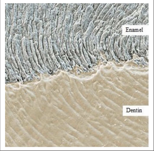
Enamel is the outermost and hardest tissue in the human body.[4] It consists of approximately 96% inorganic material, primarily in the form of carbonated hydroxyapatite crystals, with the remainder composed of organic matrix and water.[5] Its main function is to protect the underlying dentine and aid in food breakdown through mastication.[6]
Enamel is formed during amelogenesis, a two-stage process beginning with the secretion of an organic matrix by ameloblasts near the dentinoenamel junction (DEJ).[5] Once mineralisation reaches 96%, enamel formation is complete, and no further deposition occurs due to the degeneration of ameloblasts.[7][5]
At the microscopic level, enamel has a complex structure composed of enamel rods and interrod enamel, arranged in a prism-like pattern which contributes to its density and mechanical strength.[5]
Dentine
Dentine lies beneath the enamel and forms the bulk of the anatomical crown. It supports the enamel and protects the innermost pulp chamber.[7][6] Composed of 70% inorganic material, 20% organic matrix (mainly collagen), and 10% water, dentine is resilient and capable of absorbing functional stresses.[8]
The structure of dentine includes dentinal tubules that extend from the enamel-dentine junction to the pulp. These tubules are surrounded by peritubular and intertubular dentine, contributing to its mechanical properties and sensitivity.[5][8]

Pulp chamber
The pulp chamber is the innermost part of the anatomical crown and contains blood vessels, nerves, lymphatics, and odontoblasts.[3] It plays a role in dentine formation, nutrient delivery, and pain response.
Though mainly associated with root canal treatments, the pulp chamber's position within the crown is important in clinical practice. Several anatomical "laws" have been described to aid in locating the pulp chamber during restorative or endodontic procedures:
- Law of centrality: The pulp chamber is always located in the centre of the tooth at the level of the cementoenamel junction (CEJ).[10]
- Law of concentricity: At the CEJ level, the pulp chamber walls are concentric to the external surface of the crown.[10]
- Law of the CEJ: The CEJ is the most consistent landmark for locating the pulp chamber.[10]
Differences between primary and permanent crowns

Primary teeth are the first set of teeth in the human dentition. It comprises 20 teeth, known as primary teeth or milk teeth. Primary teeth begin to erupt in infancy and are eventually replaced by permanent teeth. Premature loss of primary teeth can result in malocclusion or crowding of the permanent successors.[11]
Primary teeth differ from permanent teeth in several anatomical and structural ways. The crowns of primary teeth are generally shorter and broader, with a thinner layer of enamel, making them more susceptible to wear. This enamel also gives them a whiter appearance compared to permanent teeth.[11] In anterior teeth, mamelons, small bumps on the incisal edge of newly erupted permanent incisors, are absent in the primary dentition. The cervical ridges are more pronounced, particularly in molars, and the crowns are more bulbous with a distinct cervical constriction.[11]
Structurally, the roots of primary teeth are thinner and more widely spread, with short or absent root trunks. These adaptations facilitate natural exfoliation as the underlying permanent teeth erupt.[11]
من الناحية الوظيفية والمورفولوجية، تتميز الأضراس اللبنية بسطح إطباق أضيق وسطحين شفهيين ولسانيين أكثر استواءً، بينما تكون الأسنان اللبنية الأمامية أعرض نسبيًا من الناحية الإنسية الوحشية مقارنةً بارتفاع تاجها. هذه الفروقات مهمة أثناء تقييمات الأسنان وإجراءات الترميم. [ 12 ] [ 13 ]
التباين المورفولوجي بين أنواع الأسنان
القواطع
في الأسنان الدائمة، تتميز القواطع المركزية العلوية بتيجان عريضة مستطيلة الشكل ذات حافة قاطعة مستقيمة. غالبًا ما تظهر على القواطع حديثة البزوغ ثلاثة نتوءات، تتآكل مع مرور الوقت. يحتوي السطح اللساني على حزام واضح محاط بحواف هامشية وسطية وبعيدة، ويحيط بحفرة لسانية ضحلة. [ 12 ] أما القواطع الجانبية العلوية فهي أصغر حجمًا، ذات زوايا قاطعة مستديرة وحفرة لسانية أعمق قد تحتوي على أخاديد نمو. [ 12 ] بينما تُعد القواطع المركزية السفلية أصغر الأسنان، وتتميز بتاج متناظر ذي حافة قاطعة مستقيمة وسطح لساني أملس. أما القواطع الجانبية السفلية فهي أكبر حجمًا قليلًا ولها حافة قاطعة مائلة نحو الخلف. [ 12 ] [ 14 ]
تتميز القواطع المركزية العلوية اللبنية بتيجان أعرض في الاتجاه الميزي البعيد منها في الاتجاه القاطع العنقي، وهي سمة لا توجد في أي سن آخر. وهي تفتقر إلى النتوءات المينائية، وتتميز بوجود حزام بارز وحواف هامشية مع حفرة لسانية أعمق. [ 15 ] أما القواطع الجانبية اللبنية فهي أصغر حجماً بزوايا قاطعة أكثر استدارة. في الفك السفلي، تكون القواطع المركزية اللبنية متناظرة ذات تاج مدبب وسطح لساني أملس، بينما تكون القواطع الجانبية أكبر قليلاً ذات حافة قاطعة مائلة نحو الخلف. [ 15 ]
الكلاب
تتميز الأنياب العلوية الدائمة بوجود حافة شفوية بارزة، وحزام متطور ، ونتوء مدبب واضح. يبدو تاج السن على شكل ماسة من المنظر القاطع، مع انحدارات قوية من الجهتين الإنسية والوحشية. يشمل التشريح اللساني حافة مركزية محاطة بحفر ضحلة وحواف هامشية بارزة. [ 12 ] [ 16 ] أما الأنياب السفلية فهي أضيق من الناحية الإنسية الوحشية، مع حزام أقل بروزًا وسطح لساني أكثر نعومة. وتكون تيجانها بشكل عام أكثر تسطحًا وأقل استدارة من تيجان الأنياب العلوية. [ 16 ]
في الأسنان اللبنية، تتميز الأنياب العلوية بنتوءات بارزة وحادة ذات منحدرات أمامية أطول. ويكون تاج السن ضيقًا عند العنق ويبدو أكثر انتفاخًا. [ 12 ]
الضواحك
Maxillary premolars usually have two cusps–buccal and lingual. The first premolars show sharp buccal cusp and a smaller lingual cusp separated by a central groove. They often exhibit a pronounced buccal ridge and occlusal sulcus.[12][13] The second premolars are smaller with cusps of more equal height and display more supplemental grooves on the occlusal surface.
Mandibular first premolars have a dominant buccal cusp and a much smaller lingual cusp, often giving the appearance of a single cusp. The crown tapers sharply towards the lingual side.[13] Second premolars typically have two lingual cusps and a broader, square, or round occlusal table. Their occlusal groove pattern may vary from Y, H, to U shapes.[13]
Molars
Maxillary first permanent molars have four main cusps, and sometimes a fifth cusp known as the cusp of Carabelli. The occlusal surface typically has a rhomboidal shape and includes a distinct oblique ridge connecting the mesiopalatal and distobuccal cusps.[12] Second maxillary molars are smaller, and the distopalatal cusp may be reduced or absent, giving rise to a heart-shaped occlusal form. Third molars are highly variable in anatomy, often smaller and more rounded, with numerous accessory grooves and ridges.[12]
Mandibular first molars have five cusps and a pentagonal occlusal outline. They include three buccal and two lingual cusps, separated by distinct grooves. The mesiobuccal cusp is typically the largest.[17] Second molars have four cusps of nearly equal size and a rectangular occlusal outline, with grooves forming a cross pattern. Third molars exhibit significant anatomical variation and often have wrinkled occlusal surfaces due to supplemental grooves.[12]
Primary molars differ in shape and size from permanent molars. Maxillary first molars have a prominent mesiopalatal cusp and a smooth buccal surface with minimal grooves. Second molars resemble permanent maxillary first molars and include a cusp of Carabelli.[18] Mandibular first molars are unique in shape and do not resemble any permanent teeth, featuring a strong mesial marginal ridge and pronounced curvature at the cervical third.[18] Second mandibular molars resemble the permanent mandibular first molars but are smaller in all dimensions.[18]
Radiographic appearance
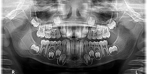
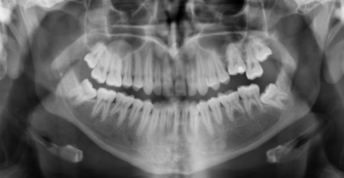
في صور الأشعة السينية، يظهر المينا كأكثر بنية معتمة للأشعة (بيضاء) نظرًا لمحتواه المعدني العالي. [ 19 ] أما العاج والملاط فهما أقل عتامة للأشعة، وعادةً ما يصعب التمييز بينهما. [ 19 ] وتكون حجرة اللب وقنوات الجذر شفافة للأشعة (داكنة)، وتقع في مركز بنية السن. ويظهر الرباط اللثوي كخط رفيع شفاف للأشعة بين الجذر والصفيحة العظمية. [ 19 ]
تشوهات النمو التي تؤثر على شكل التاج
قد تؤدي التشوهات النمائية التي تصيب تاج السن إلى تغييرات في شكله وحجمه وبنيته، مما يؤثر على مظهره ووظيفته. تنشأ هذه التشوهات عادةً خلال المراحل المبكرة من تكوين السن ، وقد ينتج عنها عيوب مثل وجود نتوءات زائدة، أو التحام الأسنان، أو انخفاضات غير طبيعية. يمكن أن تسبب هذه الاختلافات في شكل تاج السن صعوبات في الحفاظ على نظافة الفم، ومشاكل في الإطباق، وتزيد من خطر تسوس الأسنان ومشاكل اللثة.
الاندماج
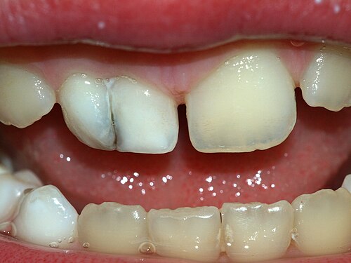
يحدث الاندماج عندما يندمج سنّان ناميان، مُشكّلين تاجًا كبيرًا قد يحتوي على جذر واحد أو جذرين. [ 21 ] يؤثر الاندماج الكامل على كلٍّ من التاج والجذور، بينما يؤثر الاندماج غير الكامل على التاج فقط. [ 22 ] قد يحتوي التاج على أخدود أو شقّ يزيد من تراكم البلاك. [ 22 ] تشمل المعالجة استخدام مواد مانعة للتسوس، أو إعادة تشكيل السن، أو خلعه إذا لزم الأمر. [ 23 ]
التوأمة
ينتج التوأمة عن محاولة سن واحد الانقسام، وتظهر على شكل تاج مشقوق بجذر واحد وقناة جذرية واحدة. [ 22 ] على عكس الاندماج، لا تؤدي التوأمة إلى تقليل عدد الأسنان. [ 23 ] تزيد الأخاديد العميقة من خطر التسوس. قد يشمل العلاج مواد مانعة للتسوس، أو ترميمات، أو تقويم الأسنان. [ 23 ]
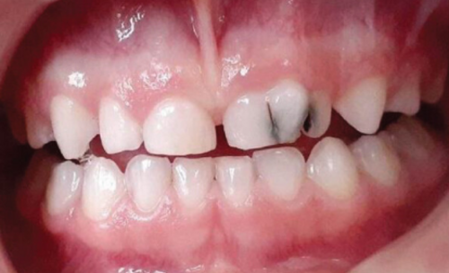
من الناحية السريرية، قد تسبب الأسنان المزدوجة مشاكل جمالية، ومشاكل في تباعد الأسنان، وسوء إطباق الأسنان. وبحسب تأثيرها، قد يشمل العلاج استخدام مواد مانعة للتسوس، أو ترميمات، أو تقويم الأسنان، أو خلع الأسنان في الحالات الشديدة. [ 23 ]
أوكار invaginatus (أوكار في دينتي)
السن المنغرز (DI)، أو السن داخل السن، هو حالة ينثني فيها جزء من تاج السن إلى الداخل قبل التصلب، أثناء النمو، مكونًا أخدودًا عميقًا أو جيبًا مبطنًا بالمينا. [ 21 ] [ 24 ] يؤثر هذا الشذوذ في أغلب الأحيان على القواطع الجانبية العلوية، تليها القواطع المركزية، ثم الضواحك. [ 24 ]
قام أوهلرز (1957) بتصنيف DI إلى ثلاثة أنواع بناءً على مدى امتداد الانغلاف داخل السن: [ 25 ]
- النوع الأول : يقتصر الانغلاف على التاج ولا يصل إلى الجذر.
- النوع الثاني : يمتد الانغلاف إلى الجذر ولكنه يبقى داخل حجرة اللب، دون أي اتصال مع اللثة.
- النوع الثالث : يمتد الانغلاف من التاج عبر الجذر، مكونًا فتحة في الرباط اللثوي إما جانبيًا (النوع الثالث أ) أو عند قمة الجذر (النوع الثالث ب).
يؤثر النوع الأول والنوع الثالث فقط بشكل مباشر على التاج، مما يؤدي إلى تغيير شكله وخصائص سطحه.
قد تبدو الأسنان المصابة على شكل برميل أو مخروطي، وهي عرضة للتسوس. [ 26 ] ويتراوح العلاج بين الحشو والخلع، وذلك حسب شدة الحالة. [ 24 ]
Dens evaginatus
يظهر السن الناشئ على شكل نتوء إضافي على السطح الإطباقي أو الحنكي، ويُعرف أيضًا باسم نتوء المخلب في الأسنان الأمامية. [ 21 ] [ 24 ] يحتوي هذا النتوء على المينا والعاج، وأحيانًا اللب. وقد يُعيق الإطباق ويزيد من خطر التسوس. [ 24 ] تشمل طرق العلاج الطحن، أو الحشو، أو علاج قناة الجذر في حال انكشاف اللب. [ 27 ] [ 28 ]
صغر الأسنان
صغر الأسنان هو خلل نمائي يظهر فيه سن واحد أو أكثر أصغر من الحجم الطبيعي، مما يؤدي غالبًا إلى مشاكل جمالية، ومشاكل في تباعد الأسنان، وصعوبة في المضغ. [ 29 ] أكثر الأسنان تأثرًا هي القواطع الجانبية العلوية، والتي غالبًا ما تظهر على شكل وتد مخروطي. [ 29 ] [ 30 ] لذلك، قد يُساهم صغر الأسنان في مشاكل وظيفية مثل انحشار الطعام بسبب عدم تباعد الأسنان بشكل صحيح، مما يؤدي إلى تسوس الأسنان ومشاكل اللثة. [ 29 ] قد يشمل العلاج تقويم الأسنان والحشوات. [ 29 ]
الأسنان الكبيرة
Macrodontia, or megalodontia, is a condition where one or more teeth are abnormally large, while still having normal crown, root, and pulp morphology.[22][30] Isolated macrodontia usually occurs in isolation, but generalised macrodontia (affecting all teeth) may be linked to systemic conditions such as otodental syndrome, insulin-resistant diabetes, and hypophyseal gigantism.[21] Not to be mistaken for gemination or fusion, macrodontia does not involve tooth splitting or merging.[22][23] Clinically, macrodontia may cause crowding, misalignment, and aesthetic concerns.[22] Treatment may include size reduction, orthodontics, or extraction.[22][30]
References
- 12345Scheid, Rickne C.; Weiss, Gabriela (2012). Woelfel's dental anatomy (8th ed.). Philadelphia: Wolters Kluwer health - Lippincott Williams & Wilkins. ISBN 978-1-60831-746-2.
- ↑Ritter, André V., ed. (2019). Sturdevant's art and science of operative dentistry (Seventh ed.). St. Louis, Missouri: Elsevier. ISBN 978-0-323-47833-5.
- 12Themes, U. F. O. (2015-01-04). "2: The Tooth: Functions and Terms". Pocket Dentistry. Retrieved 2025-04-24.
- ↑Gil-Bona, Ana; Bidlack, Felicitas B. (2020-06-23). "Tooth Enamel and Its Dynamic Protein Matrix". International Journal of Molecular Sciences. 21 (12): 4458. doi:10.3390/ijms21124458. ISSN 1422-0067. PMC 7352428. PMID 32585904.
- 12345Shahmoradi, Mahdi; Bertassoni, Luiz E.; Elfallah, Hunida M.; Swain, Michael (2014), "Fundamental Structure and Properties of Enamel, Dentin and Cementum", in Ben-Nissan, Besim (ed.), Advances in Calcium Phosphate Biomaterials, vol. 2, Berlin, Heidelberg: Springer Berlin Heidelberg, pp. 511–547, doi:10.1007/978-3-642-53980-0_17, ISBN 978-3-642-53979-4, retrieved 2025-04-24
- 12Chun, K.; Choi, H.; Lee, J. (2014-02-06). "Comparison of mechanical property and role between enamel and dentin in the human teeth". Journal of Dental Biomechanics. 5. doi:10.1177/1758736014520809. ISSN 1758-7360. PMC 3924884. PMID 24550998.
- 12Fu, Zeyu; Zhuang, Yu; Cui, Jinjie; Sheng, Ruilong; Tomás, Helena; Rodrigues, João; Zhao, Bin; Wang, Xudong; Lin, Kaili (June 2022). "Development and challenges of cells- and materials-based tooth regeneration". Engineered Regeneration. 3 (2): 163–181. doi:10.1016/j.engreg.2022.04.003.
- 12Goldberg, Michel; Kulkarni, Askok B.; Young, Marian; Boskey, Adele (2011-01-01). "Dentin: structure, composition and mineralization". Frontiers in Bioscience. 3 (2): 711–735. doi:10.2741/E281. ISSN 1945-0494. PMC 3360947. PMID 21196346.
- ↑Al-Asmar, Ayah A (February 2023). "Clinical Implementation of Comprehending Dentin on a Microscopic Level". International Journal of Morphology. 41 (1): 195–209. doi:10.4067/S0717-95022023000100195. ISSN 0717-9502.
- 123Krasner, Paul; Rankow, Henry J. (2004-01-01). "Anatomy of the Pulp-Chamber Floor". Journal of Endodontics. 30 (1): 5–16. doi:10.1097/00004770-200401000-00002. ISSN 0099-2399. PMID 14760900.
- 1234Ahmed, Nada Taha (2023). "Major contrasts between primary and permanent teeth". Research Gate.
- 12345678910Nelson, Stanley J. (2015). Wheeler's dental anatomy, physiology, and occlusion (Tenth ed.). St. Louis, Missouri: Elsevier Saunders. ISBN 978-0-323-26323-8.
- 1 2 3 4 بلاك، جي في (1897). التشريح الوصفي لأسنان الإنسان . فيلادلفيا: شركة إس إس وايت للتصنيع.
- ^ كروسيونيسكو، إيمانويلا ليديا؛ نيجروتيو، ميدا لافينيا؛ رومينو، ميهاي؛ نوفاك، أندريا كودروتا؛ موديجا، كريستينا؛ كابلار، بوريسلاف-دوشان؛ سينيسكو، كوزمين؛ بوب ، دانييلا ماريا (23/02/2023) ، “تشريح الأسنان ومورفولوجيا الأسنان الدائمة” ، الأسنان البشرية - من الوظيفة إلى الجماليات ، IntechOpen، doi : 10.5772/intechopen.110223 ، ISBN 978-1-83768-659-9تم الاطلاع عليه بتاريخ 24 أبريل 2025
- 1 2 susanhenderson (2024-06-01). "مورفولوجيا الأسنان - الأسنان اللبنية (الجزء ب)" .
{{cite journal}}يتطلب الاستشهاد بالمجلة ( مساعدة )|journal= - 1 2 فولر، جيمس ل.؛ دينيهي، جيرالد إي.؛ جيرش، ويليام (1977). تشريح وشكل الأسنان الموجز (باللغة اليابانية). دار نشر الكتاب الطبي السنوي.
- ↑ أرولا، د.؛ ريبروجيل، ر.ك. (1 يونيو 2005). "تأثيرات الشيخوخة على السلوك الميكانيكي لعاج الأسنان البشري" . المواد الحيوية . 26 (18): 4051-4061 . doi : 10.1016/j.biomaterials.2004.10.029 . ISSN 0142-9612 . PMID 15626451 .
- 1 2 3 دين، جيه إيه؛ جونز، جيه إي؛ ساندرز، بي جيه؛ فينسون، إل إيه دبليو؛ ييبس، جيه إف (2022). طب أسنان ماكدونالد وأيفري للأطفال والمراهقين ( الطبعة الحادية عشرة). إلسيفير. ISBN 978-0-323-69820-7.
- 1 2 3 نيب، هنري؛ واليزاي، طارق؛ هاكينج، كريج (13-06-2014)، "الأسنان" ، Radiopaedia.org ، doi : 10.53347/rid-29678 ، تاريخ الاسترجاع : 24-04-2025
- ↑ كيير، إنجر (2014). "آلية بزوغ الأسنان البشرية: مقالة مراجعة تتضمن نظرية جديدة للدراسات المستقبلية حول عملية البزوغ" . ساينتيفيكا . 2014 ( 1) 341905. doi : 10.1155/2014/341905 . ISSN 2090-908X . PMC 3944225. PMID 24688798 .
- 1 2 3 4 كارجودكار، إف آر؛ مالي، إس؛ سونتاكي، إس؛ سانساري، ك؛ باتيل، دي جيه (2012). "JCDR - الاندماج، والسن الزائد، وانغلاف السن، ونتوء تالون، والتمزقات" . مجلة البحوث السريرية والتشخيصية . 6 (9): 1603-1605 . doi : 10.7860/jcdr/2012/4174.2576 . PMC 3527813. PMID 23285473. تاريخ الاسترجاع : 24-04-2025 .
- ١ ٢ ٣ ٤ ٥ ٦ ٧ مامداني، ساجدة؛ باثاك، ديفيا؛ هاريسون، مايك؛ بهوجيل، نابينا (مارس ٢٠٢٣). "تضخم الأسنان والأسنان المزدوجة: مراجعة وسلسلة حالات" . المجلة البريطانية لطب الأسنان . ٢٣٤ (٥): ٣١٥-٣٢١ . doi : 10.1038/s41415-023-5571-9 . ISSN 1476-5373 . PMID 36899237 .
- ١ ٢ ٣ ٤ ٥ بن سالم، منى؛ شوشان، فرح؛ مسعودي، فاطمة؛ بعزيز، أحلام؛ معتوق، فتحي؛ غديرة، هشام (٢٠٢١). "اندماج أم توأمة؟ التشخيص والعلاج في الأسنان اللبنية: تقرير حالتين" . تقارير حالات في طب الأسنان . ٢٠٢١ (١) ٦٦٦١٧٧٦. doi : 10.1155/2021/6661776 . ISSN 2090-6455 . PMC 8189771. PMID 34123435 .
- 1 2 3 4 5 ساهني، هيمانث؛ أغاروال، فانشيكا؛ غوبتا، جيتانجالي (ديسمبر 2023). "تشخيص بروز وانغلاف السن في القاطع الجانبي العلوي الممزق باستخدام التصوير المقطعي المحوسب ذي الحزمة المخروطية - تقرير حالة ومراجعة للأدبيات" . المجلة الهندية لأبحاث طب الأسنان . 34 (4): 448-450 . doi : 10.4103/ijdr.ijdr_53_22 . ISSN 0970-9290 . PMID 38739830 .
- ↑Siqueira, José F.; Rôças, Isabela N.; Hernández, Sandra R.; Brisson-Suárez, Karen; Baasch, Alessandra C.; Pérez, Alejandro R.; Alves, Flávio R. F. (2022-02-01). "Dens Invaginatus: Clinical Implications and Antimicrobial Endodontic Treatment Considerations". Journal of Endodontics. 48 (2): 161–170. doi:10.1016/j.joen.2021.11.014. ISSN 0099-2399. PMID 34902355.
- ↑Chaturvedula, Balavenkata Bharathi; Muthukrishnan, Arvind; Bhuvaraghan, Aarthi; Sandler, Jonathan; Thiruvenkatachari, Badri (March 2021). "Dens invaginatus: a review and orthodontic implications". British Dental Journal. 230 (6): 345–350. doi:10.1038/s41415-021-2721-9. ISSN 1476-5373. PMID 33772187.
- ↑Chen, Jung-Wei; Huang, George T.-J.; Bakland, Leif K. (2020-05-01). "Dens evaginatus: Current treatment options". The Journal of the American Dental Association. 151 (5): 358–367. doi:10.1016/j.adaj.2020.01.015. ISSN 0002-8177. PMID 32209245.
- ↑Sarpangala, M.; Devasya, A. (2017). "JCDR - Accessory cusp, Mesiodens, Primary dentition". Journal of Clinical and Diagnostic Research. 11 (1): ZJ01–ZJ02. doi:10.7860/jcdr/2017/24042.9207. PMC 5324526. PMID 28274081. Retrieved 2025-04-24.
- 1234Laverty, D. P.; Thomas, M. B. M. (August 2016). "The restorative management of microdontia". British Dental Journal. 221 (4): 160–166. doi:10.1038/sj.bdj.2016.595. ISSN 1476-5373. PMID 27561572.
- 123McKinney, Reed; Brizuela, Melina; Olmo, Heather (2025), "Developmental Disturbances of the Teeth, Anomalies of Shape and Size", StatPearls, Treasure Island (FL): StatPearls Publishing, PMID 34662069, retrieved 2025-04-24
- أجزاء السن
- تشريح فم الإنسان